Enhanced Raman spectroscopy analysis for contamination detection on microelectronic devices using gold nanoclusters grown by DC magnetron sputtering
DOI:
https://doi.org/10.55713/jmmm.v33i3.1665Keywords:
Surface-enhanced Raman scattering, Sputtering, Gold nanoclusters, Failure analysisAbstract
Surface-enhanced Raman spectroscopy (SERS) is one of the most powerful analytical techniques for the identification of molecules in microelectronics industry for failure analysis protocols.
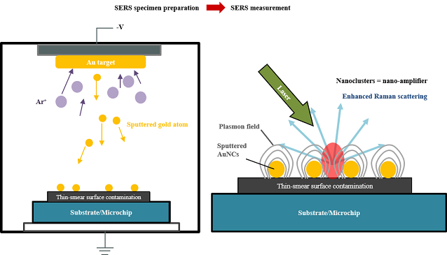
Surface-enhanced Raman spectroscopy (SERS) is one of the most powerful analytical techniques for the identification of molecules in the microelectronics industry for failure analysis protocols. In this work, dry-processed gold nanoclusters were prepared by magnetron sputtering deposition to promote the enhancement of the Raman signal from selected common polymers found in the hard disk drive as surface contamination. The optimized sputtering conditions were applied for SERS on poly-carbonate (PC), polyethylene terephthalate (PET), polypropylene (PP), and high-density polyethylene (HDPE). The Raman spectrum showed the average Raman signal intensity gain at about 114%, 78%, 254%, and 226%, respectively. The SERS with gold nanoclusters, prepared by magnetron sputtering, demonstrates that this method is a clean, simple, highly performing analytical method for failure analysis and can be an alternative method over the use of colloidal gold nanoparticles for contamination investigation in industrial failure analysis procedures, where the sample cleanness during the analysis is critical, as in the microelectronic industry.
Downloads
References
K. M. Deen, and I. H. Khan, “Chapter 2 - Engineering failure analysis in chemical process industries,” in Handbook of Materials Failure Analysis with Case Studies from the Chemicals, Concrete and Power Industries, A. S. H. Makhlouf and M. Aliofkhazraei, Eds., Butterworth-Heinemann, 2016, pp. 25-47.
L. Xia, J. Wang, S. Tong, G. Liu, J. Li, and H. Zhang, “Design and construction of a sensitive silver substrate for surface-enhanced Raman scattering spectroscopy,” Vibrational Spectroscopy, vol. 47, no. 2, pp. 124-128, 2008.
Y. Wang, H. Wei, B. Li, W. Ren, and S. Guo, S. Dong, and E. Wang, “SERS opens a new way in aptasensor for protein recognition with high sensitivity and selectivity,” Chemical Communications, vol. 0, no. 48, pp. 5220-5222, 2007.
R. Pilot, R. Signorini, C. Durante, L. Orian, M. Bhamidipati, and L. Fabris, “A Review on surface-enhanced raman scattering,” Biosensors, vol. 9, no. 2, no. 2, 2019.
M. Ocaña, V. Fornés, J. V. G. Ramos, and C. J. Serna, “Factors affecting the infrared and Raman spectra of rutile powders,” Journal of Solid State Chemistry, vol. 75, no. 2, pp. 364-372, 1988.
D. Tuschel, “Raman spectroscopy and polymorphism,”
Spectroscopy, vol. 34, no. 3, pp. 10-21, 2019.
B. Sharma, R. R. Frontiera, A.-I. Henry, E. Ringe, and R. P. V. Duyne, “SERS: Materials, applications, and the future,” Materials Today, vol. 15, no. 1, pp. 16-25, 2012.
S. M. Novikov, V. N. Popok, A. B. Evlyukhin, M. Hanif, P. Morgen, J. Fiutowski, J. Beermann, H-G Rubahn, and S. I. Bozhevolnyi, “Highly stable silver nanoparticles for SERS applications,” Journal of Physics: Conference Series, vol. 1092, p. 012098, 2018.
J. Sarkar, “Chapter 2 - Sputtering and thin film deposition,” in Sputtering Materials for VLSI and Thin Film Devices, J. Sarkar, Ed., Boston: William Andrew Publishing, 2014, pp. 93-170.
N. Guillot, and M. L. de la Chapelle, “The electromagnetic effect in surface enhanced Raman scattering: Enhancement optimization using precisely controlled nanostructures,” Journal of Quantitative Spectroscopy and Radiative Transfer, vol. 113, no. 18, pp. 2321-2333, 2012.
F. M. Liu, P. A. Köllensperger, M. Green, A. E. G. Cass, and L. F. Cohen, “A note on distance dependence in surface enhanced Raman spectroscopy,” Chemical Physics Letters, vol. 430, no. 1, pp. 173-176, 2006.
M. Pišlová, M. Kalbacova, L. Vrabcová, P. Slepička, Z. Kolská, and V. Švorčík, “Preparation of noble nanoparticles by sputtering – their characterization,” Digest Journal of Nano-materials and Biostructures, vol. 13, pp. 1035-1044, 2018.
M. Nie, K. Sun, and D. D. Meng, “Formation of metal nano-particles by short-distance sputter deposition in a reactive ion etching chamber,” Journal of Applied Physics, vol. 106, no. 5, p. 054314, 2009.
G. Zhiqi, “Raman spectroscopy analysis of minerals,” Raman spectroscopy analysis of minerals based on feature visualization [Online], Available: https://www.spectroscopyonline.com/ view/
raman-spectroscopy-analysis-of-minerals-based-on-feature-visualization
F. Adar, “Introduction to interpretation of raman spectra using database searching and functional group detection and identification” [Online], Jul 01, 2016. Available: https://www.
spectroscopyonline.com/view/introduction-interpretation raman-
spectra-using-data-base-searching-and-functional-group-detection-a
R. Wojnarowska-Nowak, J. Polit, D. Broda, Y. Bobitski, Z. Starowicz, M. Gonchar, and E. M. Sheregii, “Surface-enhanced raman scattering and plasmon effect for enzymatic bionano-complexes characterization,” EPJ Web of Conference, vol. 133, p. 05001, 2017.
E. Rebollar, S. Pérez, M. Hernandez, C. Domingo, M. Martín, T. A. Ezquerra, J. P. García-Ruiz, and M.Castillejo, “Physico-chemical modifications accompanying UV laser induced surface structures on poly(ethylene terephthalate) and their effect on adhesion of mesenchymal cells,” Physical Chemistry Chemical Physics, vol. 16, no. 33, pp. 17551-17559, 2014.
V. Dhaka, S. Singh, A. G. Anil, T. S. Sunil Kumar Naik, S. Garg, J. Samuel, M. Kumar, P. C. Ramamurthy, and J. Singh, “Occurrence, toxicity and remediation of polyethylene terephthalate plastics. A review,” Environmental Chemistry Letters, vol. 20, no. 3, pp. 1777-1800, 2022.
R. M. Khafagy, “In situ FT‐Raman spectroscopic study of the conformational changes occurring in isotactic polypropylene during its melting and crystallization processes,” Journal of Polymer Science Part B: Polymer Physics, vol. 44, pp. 2173-2182, 2006.
M. Gahleitner, and C. Paulik, “Polypropylene,” in Ullmann’s Encyclopedia of Industrial Chemistry, John Wiley & Sons, Ltd, 2014, pp. 1-44.
S. V. Boriskina, “An ode to polyethylene,” MRS Energy & Sustainability, vol. 6, no. 1, p. 14, 2019.
A. P. Kotula, M. W. Meyer, F. De Vito, J. Plog, A. R. Hight Walker,
and K. B. Migler, “The rheo-raman microscope: simultaneous chemical, conformational, mechanical, and micro-structural measures of soft materials,” Review of Scientific Instruments, vol. 87, no. 10, p. 105105, 2016.
P. M. V. Raja, and A. R. Barron, “4.3: Raman Spectroscopy,” Chemistry LibreTexts [Online], Mar 22, 2021. Available: https:// chem.libretexts.org/Bookshelves/Analytical_Chemistry/Physical_Methods_in_Chemistry_and_Nano_Science_(Barron)/04%3A_Chemical_Speciation/4.03%3A_Raman_Spectroscopy
K. M. Kosuda, J. M. Bingham, K. L. Wustholz, R. P. V. Duyne, and R. J. Groarke, “4.06 - Nanostructures and Surface-enhanced raman spectroscopy☆,” in Comprehensive Nanoscience and Nanotechnology (Second Edition), D. L. Andrews, R. H. Lipson, and T. Nann, Eds., 2nd Edition. Oxford: Academic Press, 2016, pp. 117-152.
W. E. Smith, “Practical understanding and use of surface enhanced Raman scattering/surface enhanced resonance Raman scattering in chemical and biological analysis,” Chemical Society Reviews, vol. 37, no. 5, pp. 955-964, 2008.
I. Gajdos, F. Greskovic, J. Slota, and Ľ. Dulebová, “Raman spectroscopy in polymer processing technologies,” Acta Mechanica Slovaca, vol. 15, pp. 68-73, 2011.
E. Hauser, “Polymer analysis using raman spectroscopy,” AZoM.com [Online], Aug 26, 2014. Available: https://www. azom.com/article.aspx?ArticleID=11282
U. W. Gedde, “Chain Orientation,” in Polymer Physics, U. W. Gedde, Ed., Dordrecht: Springer Netherlands, 1999, pp. 199-216.

Downloads
Published
How to Cite
Issue
Section
License
Copyright (c) 2023 Journal of Metals, Materials and Minerals

This work is licensed under a Creative Commons Attribution-NonCommercial-NoDerivatives 4.0 International License.
Authors who publish in this journal agree to the following terms:
- Authors retain copyright and grant the journal right of first publication with the work simultaneously licensed under a Creative Commons Attribution License that allows others to share the work with an acknowledgment of the work's authorship and initial publication in this journal.
- Authors are able to enter into separate, additional contractual arrangements for the non-exclusive distribution of the journal's published version of the work (e.g., post it to an institutional repository or publish it in a book), with an acknowledgment of its initial publication in this journal.


