Electrospun polyvinyl alcohol nanofibrous membranes incorporated with silver nanowires and sambiloto (andrographis paniculata nees) for wound healing applications
DOI:
https://doi.org/10.55713/jmmm.v37i1.2738คำสำคัญ:
Silver nanowires, Sambiloto, Polyvinyl alcohol, Polyvinyl pyrrolidone, Electrospinningบทคัดย่อ
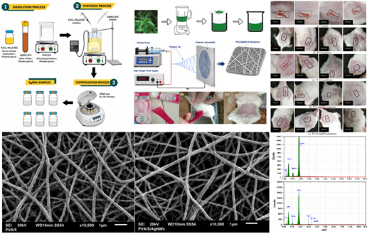
A new variation of wound covering nanofibrous membranes synthesized using electrospinning method from mixtures of polyvinyl alcohol (PVA), silver nanowires (AgNWs), and sambiloto (Andrographis paniculata Nees) leaves has successfully been developed for antibacterial applications. Four variations of nanofibrous membranes, namely pure PVA, PVA and sambiloto (PVA/S), PVA and silver nanowires (PVA/AgNWs), as well as PVA, sambiloto, and silver nanowires (PVA/S/AgNWs) were prepared and preclinically tested on white mice (Mus muscullus) that had previously been injured. The FTIR spectrum wavenumbers of PVA/S, PVA/AgNWs, and PVA/S/AgNWs for the (O-H) functional group experienced a shift greater (shifted left) than PVA. For the SEM images, the minimum diameter of the membrane nanofiber was 122 nm, while the maximum was 257 nm. The results showed that wound healing in the mice occurred after 72 h, while the PVA/S/AgNWs membranes demonstrated a more complete and accelerated wound closure than other membranes. Furthermore, the potential of sambiloto as an ingredient in a wound covering drug is relatively large. Sambiloto is an andrographolide compound used to fight disease due to its antibacterial power as well as the potential to activate lymphocytes and produce antibodies and silver has been used extensively in clinical equipment as an antimicrobial.
Downloads
เอกสารอ้างอิง
S. Guo, and L. A. DiPietro, “Factors affecting wound healing,” Journal of Dental Research, vol. 89, no. 3, pp. 219–229, 2010. DOI: https://doi.org/10.1177/0022034509359125
S. A. Eming, P. Martin, and M. Tomic-Canic, “Wound repair and regeneration: Mechanisms, signaling, and translation,” Science Translational Medicine, vol. 6, no. 265, p. 265sr6, 2014. DOI: https://doi.org/10.1126/scitranslmed.3009337
S. Dhivya, V. V. Padma, and E. Santhini, “Wound dressings—A review,” BioMedicine, vol. 5, no. 4, pp. 22–29, 2015. DOI: https://doi.org/10.7603/s40681-015-0022-9
J. S. Boateng, K. H. Matthews, H. N. E. Stevens, and G. M. Eccleston, “Wound healing dressings and drug delivery systems: A review,” Journal of Pharmaceutical Sciences, vol. 97, no. 8, pp. 2892–2923, 2008. DOI: https://doi.org/10.1002/jps.21210
G. D. Winter, “Formation of the scab and the rate of epithelization of superficial wounds in the skin of the young domestic pig,” Nature, vol. 193, pp. 293–294, 1962. DOI: https://doi.org/10.1038/193293a0
S. P. Miguel, D. R. Figueira, D. Simões, M. P. Ribeiro, P. Coutinho, P. Ferreira, and I. J. Correia, “Electrospun polymeric nanofibres as wound dressings: A review,” Colloids and Surfaces B: Biointerfaces, vol. 169, pp. 60–71, 2018. DOI: https://doi.org/10.1016/j.colsurfb.2018.05.011
S. Barrientos, O. Stojadinovic, M. S. Golinko, H. Brem, and M. Tomic-Canic, “Growth factors and cytokines in wound healing,” Wound Repair and Regeneration, vol. 16, no. 5, pp. 585–601, 2008. DOI: https://doi.org/10.1111/j.1524-475X.2008.00410.x
K. Jarukamjorn, and N. Nemoto, “Pharmacological aspects of Andrographis paniculata on health and its major diterpenoid constituent andrographolide,” Journal of Health Science, vol. 54, no. 4, pp. 370–381, 2008. DOI: https://doi.org/10.1248/jhs.54.370
P. K. Singha, S. Roy, and S. Dey, “Antimicrobial activity of Andrographis paniculata,” Fitoterapia, vol. 74, no. 7–8, pp. 692–694, 2003. DOI: https://doi.org/10.1016/S0367-326X(03)00159-X
J. Lee, Y. D. Suh, J. Lee, P. Lee, S. Han, S. Hong, J. Yeo, H. Lee, and S. H. Ko, “Recent progress in silver nanowire based flexible electrodes,” Journal of Materials Chemistry C, vol. 6, pp. 7445–7461, 2018. DOI: https://doi.org/10.1039/C8TC01024B
T. Tomiyama, I. Mukai, H. Yamazaki, and Y. Takeda, "Optical properties of silver nanowire/polymer composite films: Absorption, scattering, and color difference," Optical Materials Express, vol. 10, no. 12, pp. 3202‒3214, 2020. DOI: https://doi.org/10.1364/OME.412015
S. Hou, J. Liu, F. Shi, G.-X. Zhao, J.-W. Tan, and G. Wang, “Recent advances in silver nanowires electrodes for flexible organic/perovskite light-emitting diodes,” Frontiers in Chemistry, vol. 10, p. 864186, 2022. DOI: https://doi.org/10.3389/fchem.2022.864186
S. Chernousova, and M. Epple, “Silver as antibacterial agent: Ion, nanoparticle, and metal,” Angewandte Chemie International Edition, vol. 52, no. 6, pp. 1636–1653, 2013. DOI: https://doi.org/10.1002/anie.201205923
I. Sondi, and B. Salopek-Sondi, “Silver nanoparticles as anti-microbial agent: A case study on E. coli as a model for Gram-negative bacteria,” Journal of Colloid and Interface Science, vol. 275, no. 1, pp. 177–182, 2004. DOI: https://doi.org/10.1016/j.jcis.2004.02.012
D. H. Reneker, and I. Chun, “Nanometre diameter fibres of polymer, produced by electrospinning,” Nanotechnology, vol. 7, no. 3, pp. 216–223, 1996. DOI: https://doi.org/10.1088/0957-4484/7/3/009
S. Ramakrishna, K. Fujihara, W.-E. Teo, T. Yong, Z. Ma, and R. Ramaseshan, “Electrospun nanofibers: Solving global issues,” Materials Today, vol. 9, no. 3, pp. 40–50, 2006. DOI: https://doi.org/10.1016/S1369-7021(06)71389-X
Y. Si, X. Tang, J. Yu, and B. Ding, “Electrospun nanofibers: Solving global issues,” in Electrospun Nanofibers for Energy and Environmental Applications. Berlin, Germany: Springer, pp. 3–38, 2014. DOI: https://doi.org/10.1007/978-3-642-54160-5_1
E. A. Kamoun, S. A. Loutfy, Y. Hussein, and E.-R. Kenawy, “Recent advances in PVA-polysaccharide based hydrogels and electrospun nanofibers in biomedical applications: A review,” International Journal of Biological Macromolecules, vol. 187, pp. 755–768, 2021. DOI: https://doi.org/10.1016/j.ijbiomac.2021.08.002
Y. Yang, Z. Zhang, M. Wan, Z. Wang, X. Zou, Y. Zhao, and L. Sun, “A facile method for the fabrication of silver nanoparticles surface decorated polyvinyl alcohol electrospun nanofibers and controllable antibacterial activities,” Polymers, vol. 12, p. 2486, 2020. DOI: https://doi.org/10.3390/polym12112486
K. T. Cao, K. K. Tran, and H. T. Do, “Polyol method and surface functionalization of silver nanowires using bovine serum albumin for surface-enhanced Raman scattering application,” Journal of Metals, Materials and Minerals, vol. 33, no. 3, p. 1727, 2023. DOI: https://doi.org/10.55713/jmmm.v33i3.1727
J. Junaidi, K. Triyana, E. Suharyadi, H. Harsojo, and L. Y. L. Wu, “The roles of polyvinyl alcohol (PVA) as the capping agent on the polyol method for synthesizing silver nanowires,” Journal of Nano Research, vol. 49, pp. 174–180, 2017. DOI: https://doi.org/10.4028/www.scientific.net/JNanoR.49.174
J. Junaidi, M. W. Saputra, R. Marjunus, S. Sembiring, and S. Hadi, “The quenching and sonication effect on the mechanical strength of silver nanowires synthesized using the polyol method,” Molecules, vol. 26, p. 2167, 2021. DOI: https://doi.org/10.3390/molecules26082167
J. Junaidi, D. A. Malik, M. Rizki, I. Pratiwi, I. Karo-Karo, R. Marjunus, D. Asmi, and S. Hadi, “Synthesis of silver nitrate by evaporation chemical reduction process as potential materials for silver nanowires application,” Science and Technology Indonesia, vol. 9, no. 3, pp. 642–650, 2024. DOI: https://doi.org/10.26554/sti.2024.9.3.642-650
C.-W. Li, R. Fu, C. Yu, Z.-H. Li, H. Guan, D.-Q. Hu, D.-H. Zhao, and L.-C. Lu, “Silver nanoparticle/chitosan oligosaccharide/ poly(vinyl alcohol) nanofibers as wound dressings: A preclinical study,” International Journal of Nanomedicine, vol. 8, pp. 4131–4145, 2013. DOI: https://doi.org/10.2147/IJN.S51679
E. Rahmani, M. Pourmadadi, N. Zandi, A. Rahdar, and F. Baino, “pH-responsive PVA-based nanofibers containing GO modified with Ag nanoparticles: Physico-chemical characterization, wound dressing, and drug delivery,” Micromachines, vol. 13, no. 11, p. 1847, 2022. DOI: https://doi.org/10.3390/mi13111847
S. Hossain, Z. Urbi, H. Karuniawati, R. B. Mohiuddin, A. M. Qrimida, A. M. M. Allzrag, L. C. Ming, E. Pagano, and R. Capasso, “Andrographis paniculata (Burm. f.) Wall. ex Nees: An updated review of phytochemistry, antimicrobial pharmacology, and clinical safety and efficacy,” Life, vol. 11, no. 4, p. 348, 2021. DOI: https://doi.org/10.3390/life11040348
D. P. Gonde, B. K. Bhole, and K. S. Kakad, “Andrographolide, diterpenoid constituent of Andrographis paniculata: Review on botany, phytochemistry, molecular docking analysis, and pharmacology,” Phytochemistry, vol. 217, p. 113903, 2024. DOI: https://doi.org/10.1016/j.pharma.2023.10.001
E. S. Chauhan, K. Sharma, and R. Bist, “Andrographis paniculata: A review of its phytochemistry and pharmacological activities,” Research Journal of Pharmacy and Technology, vol. 12, no. 2, pp. 891‒900, 2019. DOI: https://doi.org/10.5958/0974-360X.2019.00153.7
A. May, Z. Kopecki, B. Carney, and A. Cowin, “Antimicrobial silver dressings: A review of emerging issues for modern wound care,” ANZ Journal of Surgery, vol. 92, no. 3, pp. 379–384, 2022. DOI: https://doi.org/10.1111/ans.17382
U. J. M. Ikezu, and S. N. Ugariogu, “Identification, quantification of phytochemicals and elemental analysis from ethanolic leaf extract of Andrographis paniculata,” South Asian Research Journal of Natural Products, vol. 6, no. 3, pp. 144–156, 2023.
S. R. Kumar, A. C. Jnanesha, S. Bharathkumar, K. Sravya, S. Venugopal, and R. K. Lal, “Unveiling the effect of foliar applied siliceous compounds on reducing seed shattering for the conservation of endangered kalmegh (Andrographis paniculata (Burm. f.) Wall. ex Nees.),” Biocatalysis and Agricultural Biotechnology, vol. 68, p. 103700, 2025. DOI: https://doi.org/10.1016/j.bcab.2025.103700
T. T. Hoang, and T. Q. Tran, “Investigating the effect of capping agent PVP on the synthesis of silver nanowires by polyol method and its application as flexible transparent conducting electrode,” VNUHCM Journal of Science and Technology Development, vol. 16, no. 4, pp. 52–60, 2013. DOI: https://doi.org/10.32508/stdj.v16i4.1596
J. Daphne, A. Francis, R. Mohanty, N. Ojha, and N. Das, “Green synthesis of antibacterial silver nanoparticles using yeast isolates and its characterization,” Research Journal of Pharmacy and Technology, vol. 11, no. 1, pp. 83–92, 2018. DOI: https://doi.org/10.5958/0974-360X.2018.00016.1
Z. Zhang, Y. Wu, Z. Wang, X. Zou, Y. Zhao, and L. Sun, “Fabrication of silver nanoparticles embedded into polyvinyl alcohol (Ag/PVA) composite nanofibrous films through electro-spinning for antibacterial and surface-enhanced Raman scattering (SERS) activities,” Materials Science and Engineering: C, vol. 69, pp. 462–469, 2016. DOI: https://doi.org/10.1016/j.msec.2016.07.015
Z. W. Abdullah, Y. Dong, N. Han, and S. Liu, “Water and gas barrier properties of polyvinyl alcohol (PVA)/starch (ST)/ glycerol (GL)/halloysite nanotube (HNT) bionanocomposite films: Experimental characterisation and modelling approach,” Composites Part B: Engineering, vol. 74, p. 107033, 2019. DOI: https://doi.org/10.1016/j.compositesb.2019.107033
L. V. Hai, L. Zhai, H. C. Kim, J. W. Kim, and J. Kim, “Green nanocomposites made with polyvinyl alcohol and cellulose nanofibers isolated from recycled paper,” Journal of Renewable Materials, vol. 7, no. 7, pp. 621–629, 2019. DOI: https://doi.org/10.32604/jrm.2019.06466
C. J. Tan, and Y. W. Tong, “The effect of protein structural conformation on nanoparticle molecular imprinting of ribonuclease A using miniemulsion polymerization,” Langmuir, vol. 23, pp. 2722–2730, 2007. DOI: https://doi.org/10.1021/la062178q
J. M. Yang, W. Y. Su, T. L. Leu, and M. C. Yang, “Evaluation of chitosan/PVA blended hydrogel membranes,” Journal of Membrane Science, vol. 236, pp. 39–51, 2004. DOI: https://doi.org/10.1016/j.memsci.2004.02.005
K. Qiu, and A. N. Netravali, “Halloysite nanotube reinforced biodegradable nanocomposites using noncrosslinked and malonic acid crosslinked polyvinyl alcohol,” Polymer Composites, vol. 34, pp. 799–809, 2013. DOI: https://doi.org/10.1002/pc.22482
K. Song, Y. Zhang, J. Meng, E. C. Green, N. Tajaddod, H. Li, and M. L. Minus, “Structural polymer-based carbon nanotube composite fibers: Understanding the processing–structure–performance relationship,” Materials, vol. 6, pp. 2543–2577, 2013. DOI: https://doi.org/10.3390/ma6062543
T. S. Gaaz, A. B. Sulong, M. N. Akhtar, A. A. H. Kadhum, A. B. Mohamad, and A. A. Al-Amiery, “Properties and applications of polyvinyl alcohol, halloysite nanotubes and their nanocomposites,” Molecules, vol. 20, no. 12, pp. 22833–22847, 2015. DOI: https://doi.org/10.3390/molecules201219884
P. Wang, S. Jiang, Y. Li, Q. Luo, J. Lin, L. Hu, L. Liu, and F. Xue, “Virus-like mesoporous silica-coated plasmonic Ag nanocube with strong bacteria adhesion for diabetic wound ulcer healing,” Nanomedicine: Nanotechnology, Biology and Medicine, vol. 24, p. 102381, 2021. DOI: https://doi.org/10.1016/j.nano.2021.102381
T. Jayakumar, C. Y. Hsieh, J. J. Lee, and J. R. Sheu, “Experimental and clinical pharmacology of Andrographis paniculata and its major bioactive phytoconstituent andrographolide,” Evidence-Based Complementary and Alternative Medicine, p. 84670, 2013. DOI: https://doi.org/10.1155/2013/846740
H. Suryanto, M. Muhajir, B. D. Susilo, Y. R. J. Pradana, H. W. Wijaya, A. S. Ansari, and U. Yanuhar, “Nanofibrillation of bacterial cellulose using high-pressure homogenization and its films characteristics,” Journal of Renewable Materials, vol. 9, no. 10, pp. 1717–1728, 2021. DOI: https://doi.org/10.32604/jrm.2021.015312
I. K. Dewi, T. Lestari, and S. N. Rofi’ah, “Formulation and physical test of ethanolic extract sambiloto leaves (Andrographis paniculata) ointment,” Sains Medika, vol. 6, no. 2, pp. 56–60, 2015. DOI: https://doi.org/10.26532/sainsmed.v6i2.603
M. Mishra, H. Kumar, and K. Tripathi, “Diabetic delayed wound healing and the role of silver nanoparticles,” Digest Journal of Nanomaterials and Biostructures, vol. 3, no. 2, pp. 49–54, 2008.
G. K. Devi, P. Suruthi, R. Veerakumar, S. Vinoth, R. Subbaiya, and S. Chozhavendhan, “A review on metallic gold and silver nanoparticles,” Research Journal of Pharmacy and Technology, vol. 12, no. 2, pp. 935–943, 2019. DOI: https://doi.org/10.5958/0974-360X.2019.00158.6
J. Jain, S. Arora, J. M. Rajwade, P. Omray, S. Khandelwal, and K. M. Paknikar, “Silver nanoparticles in therapeutics: Development of an antimicrobial gel formulation for topical use,” Molecular Pharmaceutics, vol. 6, no. 5, pp. 1388–1401, 2009. DOI: https://doi.org/10.1021/mp900056g
B. K. Priyanka, R. P. Ritesh, V. B. Pramod, P. C. Pavan, and N. S. Vinayak, “Silver nanoparticles—A review,” Research Journal of Pharmacy and Technology, vol. 10, no. 6, pp. 1820–1833, 2017. DOI: https://doi.org/10.5958/0974-360X.2017.00321.3
F. Paladini, and M. Pollini, “Antimicrobial silver nanoparticles for wound healing application: Progress and future trends,” Materials, vol. 12, no. 16, p. 2543, 2019. DOI: https://doi.org/10.3390/ma12162540
S. Maheshwari, R. K. Tiwari, and L. Singh, “Green expertise: Synthesis of silver nanoparticles for wound healing application—An overview,” Research Journal of Pharmacy and Technology, vol. 14, no. 2, pp. 1149–1154, 2021. DOI: https://doi.org/10.5958/0974-360X.2021.00206.7
N. H. Hoang, V. N. Nguyen, T. T. Nguyen, and T. M. D. Nguyen, “Impact of microwave synthesis time on the shape of silver nanostructures and their antibacterial activity,” Journal of Metals, Materials and Minerals, vol. 33, no. 1, pp. 101–106, 2023. DOI: https://doi.org/10.55713/jmmm.v33i1.1631
Z. Gao, S. Liu, S. Li, X. Shao, P. Zhang, and Q. Yao, “Fabrication and properties of the multifunctional rapid wound healing Panax notoginseng@Ag electrospun fiber membrane,” Molecules, vol. 28, no. 7, Art. no. 2972, 2023. DOI: https://doi.org/10.3390/molecules28072972
J.-P. Chen, and Y. Chiang, “Bioactive electrospun silver nano-particles-containing polyurethane nanofibers as wound dressings,” Journal of Nanoscience and Nanotechnology, vol. 10, no. 11, pp. 7560–7564, 2010. DOI: https://doi.org/10.1166/jnn.2010.2829
T. Toporcer, T. Grendel, B. Vidinskỳ, P. Gál, J. Sabo, and R. Hudák, “Mechanical properties of skin wounds after Atropa belladonna application in rats,” Journal of Metals, Materials and Minerals, vol. 16, no. 1, pp.25‒29, 2017.
Y. Wang, G. Yue, H. Lu, Z. Liu, N. Wang, F. Liu, H. Yin, and Y. Zhao, “Wound dressing applications of electrospun nanofibers: mechanism, construction and recent progress,” Nanoscale, vol. 18, no. 12, pp. 6203–6220, 2026. DOI: https://doi.org/10.1039/D5NR04448K
R. S. Jones, R. R. Draheim, and M. Roldo, “Silver nanowires: Synthesis, antibacterial activity and biomedical applications,” Applied Sciences, vol. 8, no. 5, p. 673, 2018. DOI: https://doi.org/10.3390/app8050673
T. M. S. Vidhya and Geetha, “In vitro evaluation of anti-inflammatory activity of Andrographis paniculata,” Research Journal of Pharmacy and Technology, vol. 11, no. 3, pp. 957–958, 2018. DOI: https://doi.org/10.5958/0974-360X.2018.00178.6
L. L. Aver'ianova, “Effect of antibiotics on the resistance of white mice to infection caused by antibiotic-resistant Staphylo-coccus strains,” Antibiotiki, vol. 21, no. 8, pp. 735‒742, 1976.
B. Reidy, A. Haase, A. Luch, K. A. Dawson, and I. Lynch, “Mechanisms of silver nanoparticle release, transformation and toxicity: A critical review of current knowledge and recommendations for future studies and applications,” Materials, vol. 6, no. 6, pp. 2295–2350, 2013. DOI: https://doi.org/10.3390/ma6062295
R. N. Mardiana, and Handayani, “Antibacterial activity of the sambiloto leaf extracts (Andrographis paniculata) to Bacillus cereus and Pseudomonas aeruginosa,” Biofarmasi, vol. 14, no. 1, pp. 19–24, 2016. DOI: https://doi.org/10.13057/biofar/f140103
J. Pusmarani, C. Dewi, J. P. Risky, H. A. H. Nur, I. F. Sasri, H. Febrianti, P. Setyo, and I. Zullies, “In vitro and in vivo study of gastroprotective effect of purified extract Andrographis paniculata,” Research Journal of Pharmacy and Technology, vol. 16, no. 8, pp. 3615–3621, 2023. DOI: https://doi.org/10.52711/0974-360X.2023.00596
N. W. S. D. Arini, B. K. Satriyasa, I. M. Jawi, and A. W. Indrayani, “Comparison of antibacterial activity of sambiloto (Andrographis paniculata) ethanol and water stem extract against methicillin-resistant Staphylococcus aureus (MRSA) ATCC3351 in vitro,” Jurnal Epidemiologi Kesehatan Komunitas, vol. 5, no. 2, pp. 69‒76, 2020. DOI: https://doi.org/10.14710/jekk.v5i2.6908
ดาวน์โหลด
เผยแพร่แล้ว
วิธีการอ้างอิง
ฉบับ
บท
Categories
การอนุญาต
ลิขสิทธิ์ (c) 2026 วารสารโลหะ, วัสดุ และแร่

This work is licensed under a Creative Commons Attribution-NonCommercial-NoDerivatives 4.0 International License.
Authors who publish in this journal agree to the following terms:
- Authors retain copyright and grant the journal right of first publication with the work simultaneously licensed under a Creative Commons Attribution License that allows others to share the work with an acknowledgment of the work's authorship and initial publication in this journal.
- Authors are able to enter into separate, additional contractual arrangements for the non-exclusive distribution of the journal's published version of the work (e.g., post it to an institutional repository or publish it in a book), with an acknowledgment of its initial publication in this journal.