
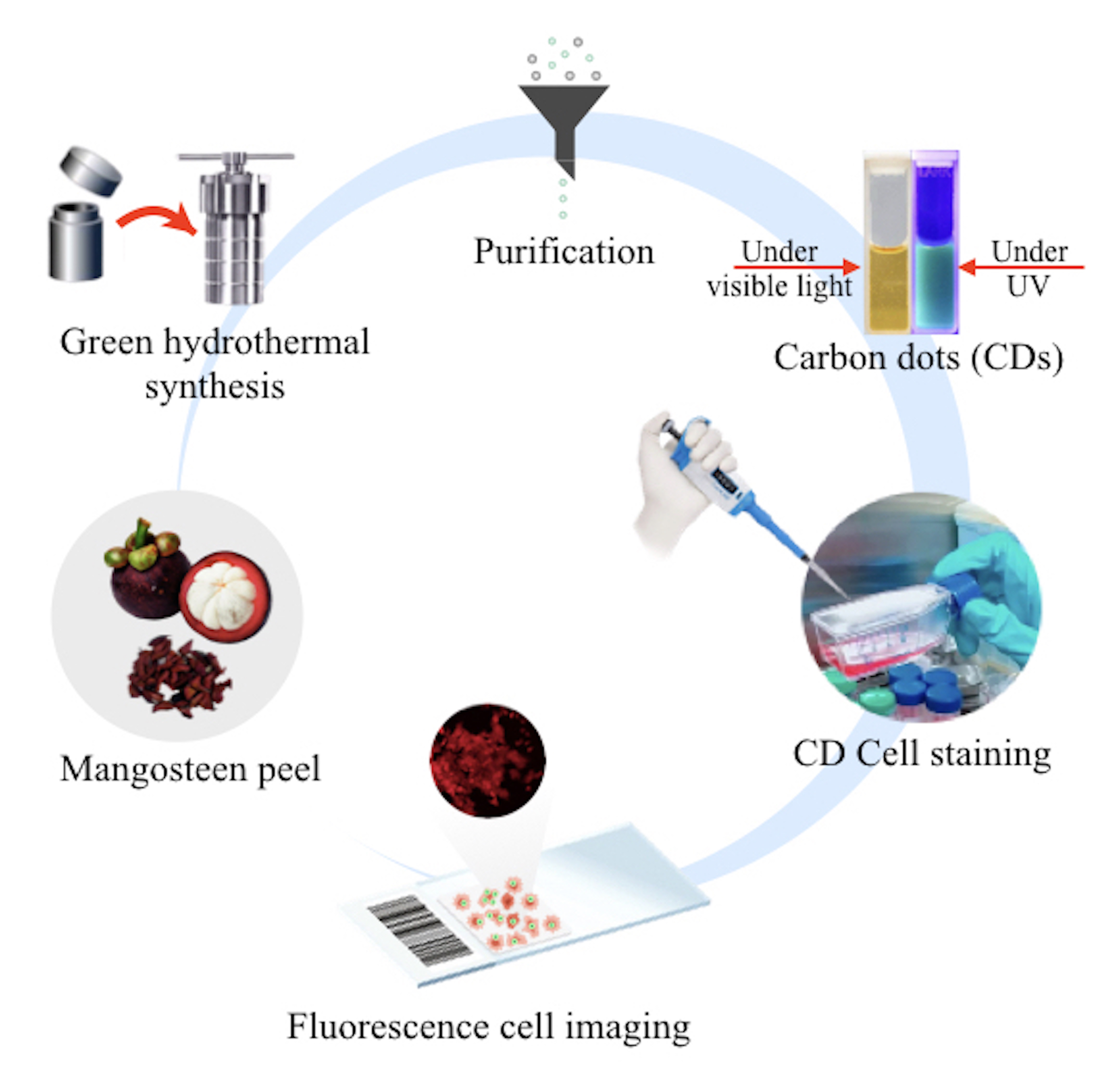
Green synthesis of carbon dots from mangosteen peel for fluorescent cancer cells
DOI:
https://doi.org/10.55713/jmmm.v34i2.1957Keywords:
Carbon dots, Mangosteen peel, Fluorescence, Caco-2 cellsAbstract
Recently, carbon dots (CDs) have received significant attention owing to their outstanding optical properties, good solubility, and low toxicity. In this research, CDs were synthesized by a hydrothermal method based on an environmentally friendly and straightforward strategy, using only mangosteen peel and deionized water. The synthesized CDs had an average size of 3.09 ± 0.38 nm. The absorbance spectrum peak for the CDs was seen at 282 nm, and the central wavelength of fluorescence emission was observed at 433 nm under an excitation wavelength of 355 nm. An aqueous solution of CDs exhibited bright green fluorescence when observed with the naked eye under UV irradiation. Both Fourier transform infrared and X-ray photoelectron spectroscope measurements were taken to determine the elemental compositions of the organic substance functional groups on the surface of the CD, such as hydroxyl, carboxyl, and carbonyl groups. These functional groups originate the different emission centers leading to multicolor fluorescent emissions. Furthermore, the synthesized CDs were found to have good biocompatibility with organic and biological materials. The remarkable properties of CDs, including their nanoscale dimensions, strong multicolor fluorescent emissions, non-toxicity, and excellent cell compatibility, could effectively permeate the cell membrane, cytoplasm, and nucleus and provide fluorescence emission. This suggests a significant potential for CDs in fluorescent cell staining applications. Finally, the CDs were used as a fluorescent dye for human colon cancer cells, as they exhibited excellent fluorescence for cell staining.
Downloads
References
M. Bruchez Jr., M. Moronne, P. Gin, S. Weiss, and A. P. Alivisatos, “Semiconductor nanocrystals as fluorescent biological labels,” Science, vol. 281, pp. 2013-2016, 1998. DOI: https://doi.org/10.1126/science.281.5385.2013
N. Thondavada, R. Chokkareddy, N. V. Naidu, and G. G. Redhi, “New generation quantum dots as contrast agent in imaging,” Nanomaterials in Diagnostic Tools and Devices, Elsevier, pp. 417-437, 2020. DOI: https://doi.org/10.1016/B978-0-12-817923-9.00015-8
S. G. Mitra, D. R. Diercks, N. C. Mills, D. L. Hynds, and S. Ghosh, “Excellent biocompatibility of semiconductor quantum dots encased in multifunctional poly (N-isopropylacrylamide) nanoreservoirs and nuclear specific labeling of growing neurons,” Applied Physics Letters, vol. 98, pp. 103702, 2011. DOI: https://doi.org/10.1063/1.3562036
G. Aizik, N.Waiskopf, M. Agbaria, Y. Levi-Kalisman, U. Banin,
and G. Golomb, “Delivery of liposomal quantum dots via monocytes for imaging of inflamed tissue,”, ACS Nano, vol. 11, pp. 3038-3051, 2017. DOI: https://doi.org/10.1021/acsnano.7b00016
C. Dias, N. Vasimalai, M. P. Sárria, I. Pinheiro, V. Vilas-Boas, J. Peixoto, and B. Espiña, “Biocompatibility and bioimaging potential of fruit-based carbon dots,” Nanomaterials, vol. 9, no. 199, pp. 1-19, 2019. DOI: https://doi.org/10.3390/nano9020199
S. Wahyudi, A. Bahtiar, C. Panatarani, Anas, and Risdiana, “Recent advanced carbon dots derived natural products and aptasensor-based carbon dots for detection of pesticides,” Sensing and Bio-Sensing Research, vol. 41, pp. 100576, 2023. DOI: https://doi.org/10.1016/j.sbsr.2023.100576
J. Liu, R. Li, and B. Yang, “Carbon dots: A new type of carbon-based nanomaterial with wide applications,” ACS Central Science, vol. 6, pp. 2179-2195, 2020. DOI: https://doi.org/10.1021/acscentsci.0c01306
N. Alvandi, S. Assariha, N. Esfandiari, and R. Jafari “Off-on sensor based on concentration-dependent multicolor fluorescent carbon dots for detecting pesticides,” Nano-Structures & Nano-Objects, vol. 26, pp. 100706, 2021. DOI: https://doi.org/10.1016/j.nanoso.2021.100706
C. E. S. Barbosa, J. R. Correa, G. A. Medeiros, G. Barreto, K. G. Magalhaes, A. L. Oliveira, J. Spencer, M. O. Rodrigues, and B. A. D. Neto, “Carbon dots (C-dots) from cow manure with impressive subcellular selectivity tuned by simple chemical modification,” Chemistry A European Journal, vol. 21, pp. 5055-5060, 2015. DOI: https://doi.org/10.1002/chem.201406330
W. Kong, J. Liu, R. Liu, H. Li, Y. Liu, H. Huang, K. Li, J. Liu, S.T. Lee, and Z. Kang, “Quantitative and real-time effects of carbon quantum dots on single living HeLa cell mem-brane permeability,” Nanoscale, vol. 6, pp. 5116, 2014. DOI: https://doi.org/10.1039/c3nr06590a
X. Gong, Q. Zhang, Y. Gao, S. Shuang, M. M. F. Choi, and C. Dong, “Phosphorus and nitrogen dual-doped hollow carbon dot as a nanocarrier for doxorubicin delivery and biological imaging,” ACS Applied Materials & Interfaces, vol. 8, pp. 11288-11297, 2016. DOI: https://doi.org/10.1021/acsami.6b01577
Y. Jiao, X. Gong, H. Han, Y. Gao, W. Lu, Y. Liu, M. Xian, S. Shuang, and C. Dong, “Facile synthesis of orange fluorescence carbon dots with excitation independent emission for pH sensing and cellular imaging,” Analytica Chimica Acta, vol 1042, pp. 125-132, 2018. DOI: https://doi.org/10.1016/j.aca.2018.08.044
J. Yang, G. Gao, X. Zhang, Y. H. Ma, X. Chen, and F. G. Wu, “One-step synthesis of carbon dots with bacterial contact-enhanced fluorescence emission: Fast Gram-type identification and selective Gram-positive bacterial inactivation,” Carbon, vol. 146, pp. 827-839, 2019. DOI: https://doi.org/10.1016/j.carbon.2019.02.040
Q. Zhang, R. Wang, B. Feng, X. Zhong, and K. Ostrikov, “Photoluminescence mechanism of carbon dots: Triggering high-color-purity red fluorescence emission through edge amino protonation,” Nature Communications, vol. 12, no. 6856, pp. 1-13, 2021. DOI: https://doi.org/10.1038/s41467-021-27071-4
V. N. Mehta, S. Jha, H. Basu, R.K. Singhal, and S. K. Kailasa, “One-step hydrothermal approach to fabricate carbon dots from apple juice for imaging of mycobacterium and fungal cells,” Sensors Actuators B: Chemical, vol. 213, pp. 434-443, 2015. DOI: https://doi.org/10.1016/j.snb.2015.02.104
R. Atchudan, T. Edison, and Y.R. Lee, “Nitrogen-doped carbon dots originating from unripe peach for fluorescent bioimaging and electrocatalytic oxygen reduction reaction,” Journal of Colloid and Interface Science, vol. 482, pp. 8-18, 2016. DOI: https://doi.org/10.1016/j.jcis.2016.07.058
A. A. Ensafi, S. S. Hghighat, N. Kazemifard, B. Rezaei, and F. Moradi, “A novel one-step and green synthesis of highly fluorescent carbon dots from saffron for cell imaging and sensing of prilocaine,” Sensor and Actuators B: Chemical, vol. 253, pp. 451-460, 2017. DOI: https://doi.org/10.1016/j.snb.2017.06.163
R. Yang, X. Guo, L. Jia, Y. Zhang, Z. Zhao, and F. Lonshakov, “Green preparation of carbon dots with mangosteen pulp for the selective detection of Fe3+ ions and cell imaging,” Applied Surface Science, vol. 423, pp. 426-432, 2017. DOI: https://doi.org/10.1016/j.apsusc.2017.05.252
R. Meena, R. Singh, G. Marappan, G. Kushwaha, N. Gupta, R. Meena, J. P. Gupta, R. R. Agarwal, N. Fahmi, and O. S. Kushwaha, “Fluorescent carbon dots driven from ayurvedic medicinal plants for cancer cell imaging and phototherapy,” Heliyon, vol. 5, pp. e02483, 2019. DOI: https://doi.org/10.1016/j.heliyon.2019.e02483
X. Y. Jiao, L. S. Li, S. Qin, Y. Zhang, K. Huang, and L. Xu, “The synthesis of fluorescent carbon dots from mango peel and their multiple applications,” Colloids and Surfaces A: Physico-chemical and Engineering Aspects, vol. 577, pp. 306-314, 2019. DOI: https://doi.org/10.1016/j.colsurfa.2019.05.073
H. Ma, C. Sun, G. Xue, G. Wu, X. Zhang, X. Han, X. Qi, X. Lv, H. Sun, and J. Zhang, “Facile synthesis of fluorescent carbon dots from Prunus cerasifera fruits for fluorescent ink, Fe3+ ion detection and cell imaging,” Spectrochimca Acta A: Molecular Biomolecular Spectroscopy, vol. 213, pp. 281-287, 2019. DOI: https://doi.org/10.1016/j.saa.2019.01.079
K. Huang, Q. He, R. Sun, L. Fang, H. Song, L. Li, Z. Li, Y. Tian, H. Cui, and J. Zhang, “Preparation and application of carbon dots derived from cherry blossom flowers,” Chemical Physics Letter, vol. 731, p. 136586, 2019. DOI: https://doi.org/10.1016/j.cplett.2019.07.014
C. Ji, Y. Zhou, R. M. Leblanc, and Z. Peng, “Recent developments of carbon dots in biosensing: A review,” ACS Sensors, vol. 5, pp. 2724-2741, 2020. DOI: https://doi.org/10.1021/acssensors.0c01556
A. A. Ridha, P. Pakravan, A. H. Azandaryani, and H. Zhaleh, “Carbon dots: The smallest photoresponsive structure of carbon in advanced drug targeting,” Journal of Drug Delivery Science and Technology, vol. 55, pp. 101408, 2019. DOI: https://doi.org/10.1016/j.jddst.2019.101408
K. A. S. Fernando, S. P. Sahu, Y. Liu, W. K. Lewis, E. Guliants, A. Jafariyan, P. Wang, C. E. Bunker, and Y. P. Sun, “Carbon quantum dots and applications in photocatalytic energy conversion,” ACS Applied Materials & Interfaces, vol.7, pp. 8363-8376, 2015. DOI: https://doi.org/10.1021/acsami.5b00448
R. Atchudan, T. N. J. I. Edison, S. Perumal, and Y. R. Lee, “Indian gooseberry-derived tunable fluorescent carbon dots as a promise for in vitro/in vivo multicolor bioimaging and fluorescent ink,” ACS Omega, vol. 3, pp. 17590-17601, 2018. DOI: https://doi.org/10.1021/acsomega.8b02463
D. Carolan, C. Rocks, D. B. Padmanaban, P. Maguire, V. Svrcek, and D. Mariotti, “Environmentally friendly nitrogen-doped carbon quantum dots for next generation solar cells,” Sustainable Energy Fuels, vol. 1, pp. 1611-1619, 2017. DOI: https://doi.org/10.1039/C7SE00158D
X. Da, Z. Han, Z. Yang, D. Zhang, R. Hong, C. Tao, H. Lin, and Y. Huang, “Preparation of multicolor carbon dots with high fluorescence quantum yield and application in white LED,” Chemical Physics Letters, vol. 794, pp. 139497, 2022. DOI: https://doi.org/10.1016/j.cplett.2022.139497
Q. Liu, S. Xu, C. Niu, M. Li, D. He, Z. Lu, L. Ma, N. Na, F. Huang, H. Jiang, and J. Ouyang, “Distinguish cancer cells based on targeting turn-on fluorescence imaging by folate functionalized green emitting carbon dots,” Biosensors and Bioelectronics, vol. 64, pp. 119-125, 2015. DOI: https://doi.org/10.1016/j.bios.2014.08.052
M. P. Aji, Susanto, P. A. Wiguna, and Sulhadi, “Facile synthesis of luminescent carbon dots from mangosteen peel by pyrolysis method,” Journal of Theoretical and Applied Physics, vol. 11, pp. 119-126, 2017. DOI: https://doi.org/10.1007/s40094-017-0250-3
A. Sangjan, S. Boonsith, K. Sansanaphongpricha, T. Thinbanmai, S. Ratchahat, N. Laosiripojana, K. C.W. Wu, H. S. Shin, and C. Sakdaronnarong, “Facile preparation of aqueous soluble fuorescent polyethylene glycol functionalized carbon dots from palm waste by one pot hydrothermal carbonization for colon cancer nanotheranostics,” Scientific Reports, vol. 12, pp. 10550, 2022. DOI: https://doi.org/10.1038/s41598-022-14704-x
P. Pakorn, S. Sangnuy, and S. Amloy, “Paper-based colorimetric sensor for mercury ion detection using smartphone digital imaging,” Journal of Metals, Materials and Minerals, vol. 33, no. 2, pp. 81-87, 2023. DOI: https://doi.org/10.55713/jmmm.v33i2.1653
D. Li, M. B. Müller, S. Gilje, R. B. Kaner, and G. G. Wallace, “Processable aqueous dispersion of graphene nanosheets,” Nature Nanotechnology, vol.3, pp. 101-105, 2008. DOI: https://doi.org/10.1038/nnano.2007.451
N. Sharma, I. Sharma, and M. K. Bera, “Microwave-assisted green synthesis of carbon quantum dots derived from Calotropis gigantea as a fluorescent probe for bioimaging,” Journal of Fluorescence, vol.32, pp. 1039-1049, 2022 DOI: https://doi.org/10.1007/s10895-022-02923-4
H. Lia, S. Hana, B. Lyua, T. Hong, S. Zhia, L. Xuc, F. Xued, L. Saie, J. Yanga, X. Wanga, and B. Heb, “Tunable light emission from carbon dots by controlling surface defects,” Chinese Chemical Letters, vol. 32, pp. 2887-2892, 2021. DOI: https://doi.org/10.1016/j.cclet.2021.03.051
Downloads
Published
How to Cite
Issue
Section
License
Copyright (c) 2024 Journal of Metals, Materials and Minerals

This work is licensed under a Creative Commons Attribution-NonCommercial-NoDerivatives 4.0 International License.
Authors who publish in this journal agree to the following terms:
- Authors retain copyright and grant the journal right of first publication with the work simultaneously licensed under a Creative Commons Attribution License that allows others to share the work with an acknowledgment of the work's authorship and initial publication in this journal.
- Authors are able to enter into separate, additional contractual arrangements for the non-exclusive distribution of the journal's published version of the work (e.g., post it to an institutional repository or publish it in a book), with an acknowledgment of its initial publication in this journal.